The Ophthalmic Electrophysiology System is a sophisticated diagnostic device that provides objective, functional assessment of the entire visual pathway by measuring the electrical signals generated by the retina and brain in response to visual stimuli.
A crucial instrument for diagnosing functional disorders often undetectable by structural imaging (like OCT or Fundus Photography), Electrophysiology is essential for localizing pathology along the visual pathway. The system performs several key tests, including: Electroretinogram (ERG), which measures retinal response; Electro-oculogram (EOG), which assesses the function of the retinal pigment epithelium; and Visual Evoked Potentials (VEP), which assesses the function of the optic nerve and visual cortex. This objective data is highly valued by eye care professionals, particularly in diagnosing inherited retinal diseases, optic neuropathies, and drug toxicities.
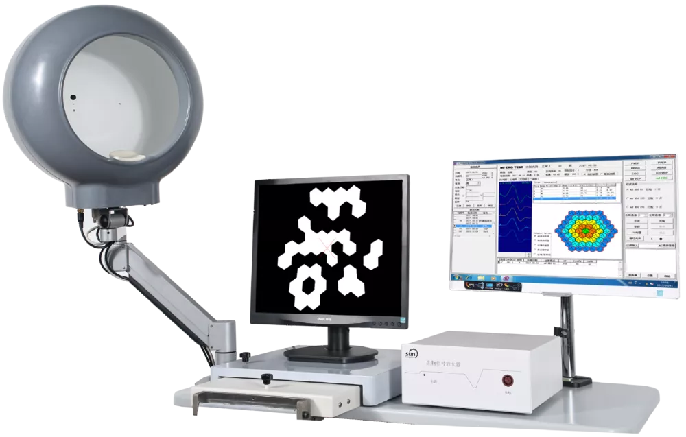
The system utilizes a specialized light source (often a Ganzfeld dome or pattern monitor) to deliver standardized visual stimuli, while small skin or corneal electrodes record the resulting electrical activity. The software processes and amplifies these minute signals to provide quantitative, color-coded waveform analysis compared against reference data. This capability makes it an indispensable tool in large referral centers, university hospitals, and specialized neuro-ophthalmology clinics.
Because of its ability to provide quantifiable and repeatable data on functional health, the Electrophysiology System is crucial for differential diagnosis, monitoring disease progression (especially in glaucoma and macular diseases), and evaluating the efficacy and safety of new treatments. Its vital role ensures confident clinical decisions in complex cases, guiding treatment and preserving long-term visual potential.