The Ophthalmic Ultrasound System is an invaluable diagnostic tool that uses high-frequency sound waves to generate detailed images of the eye's internal structures, particularly when optical viewing is obscured by conditions like dense cataracts or vitreous hemorrhage.
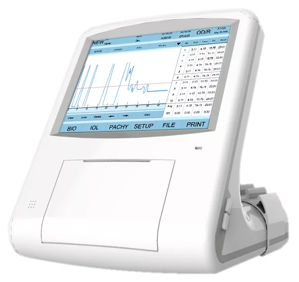
A fundamental instrument for imaging the posterior segment when opacities prevent direct visualization (e.g., via slit lamp or fundus camera), Ultrasound is essential for assessing the retina, choroid, and vitreous. The system typically performs two primary functions: B-Scan (two-dimensional imaging) for visualizing pathology like retinal detachments and intraocular tumors, and A-Scan (one-dimensional biometry) for precise measurement of axial length, which is crucial for IOL power calculation before cataract surgery.
The system features a high-frequency probe placed gently on the closed eyelid (B-Scan) or directly on the anesthetized cornea (A-Scan). This non-invasive, quick procedure produces real-time, cross-sectional (B-Scan) or quantitative (A-Scan) data. This capability simplifies the pre-surgical assessment of candidates with dense opacities, making it an indispensable tool in surgical centers and vitreoretinal practices.
Because of its ability to provide objective structural information regardless of media clarity, the Ophthalmic Ultrasound System is crucial for diagnosing, measuring, and planning treatment for retinal detachment, vitreous hemorrhage, and ocular masses. Its vital role ensures confident surgical and therapeutic decisions, leading to optimal visual outcomes.